


New Discovery: Development of the Inner Ear in Embryos is Similar to Crystal Formation
This discovery could contribute to the development of treatments for hearing loss based on regeneration of hair cells in the ear.
An interdisciplinary study headed by Prof. David Sprinzak, a researcher from the School of Neurobiology, Biochemistry and Biophysics at the George S. Wise Faculty of Life Sciences at Tel Aviv University, showed for the first time that physical forces are involved in the development of the inner ear in mammalian embryos.
Prof. Sprinzak: “We identified a new developmental mechanism that is driven by mechanical forces: the organization of hair cells in the inner ear resembles the way atoms are organized into a crystal. This is a revolutionary finding that changes fundamental perceptions in the field of developmental biology.”
The study was performed by Roie Cohen and Liat Amir-Zilberstein of Prof. Sprinzak’s laboratory; Prof. Karen Avraham and Shahar Taiber of the Sackler Faculty of Medicine; and other researchers from the Faculty of Exact Sciences and from the Sagol School of Neuroscience at Tel Aviv University. Researchers from Switzerland, and Japan also participated in the study. The paper describing the work was published in the prestigious journal, Nature Communications, in October 2020.
Prof. Sprinzak explains: “The mammalian ear has three parts: the outer, middle, and the inner ear. Within the inner ear, there is a spiral structure, the cochlea, which contains sensory cells called hair cells. Hearing occurs when sound waves entering the inner ear cause the tiny hairs located on the hair cells to vibrate. These vibrations are then converted into electrical signals that are transmitted to the brain.
The hair cells in the cochlea are arranged in a highly organized pattern, where the hair cells and a second cell type called supporting cells form an alternating checkerboard-like pattern. This organization is important since different regions along the cochlea respond to different sound frequencies. Such a remarkably organized cellular system is rather uncommon in nature – in fact, the inner ear is one of the most organized tissues in the mammalian body. In this work, we investigated the mechanism that causes the hair cells to arrange in such a pattern during embryonic development. To do so, we conducted an interdisciplinary study that involved two innovative approaches: a new imaging technology and computational simulations of the process.”
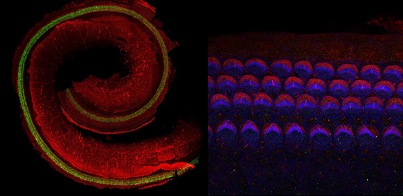
In order to track the development of hair cells in the embryo, the researchers studied mouse embryos at different developmental stages. They found that early on, the cells in the tissue are disordered and undifferentiated; namely, their type and position was not determined yet. As development progresses, the cells differentiate into hair cells and supporting cells. Then, they gradually rearrange into the organized checkboard-like pattern.
Prof. Sprinzak: “Until now, most researchers in the field focused on the process of cell differentiation, which is controlled by intercellular communication. We hypothesized that this was not enough to explain the observed behavior, and we decided to examine how cells rearrange to form an ordered pattern after differentiation.” In order to do so, the researchers developed a new imaging technology that is based on three-dimensional time lapse imaging of the inner ear using a specialized microscopy setup. This approach allowed generating time-lapse videos of the development of the tissue and to track the morphological processes occurring over several days.
Prof. Sprinzak: “This is the first time that the process has been observed continuously and at high resolution. We observed that the initially disordered hair cells and supporting cells actively move until they gradually arrange into an ordered array. Neighboring cells, known as Hensen cells, move in one direction, exerting shear forces on the hair cells – forces that act in parallel to the layer of cells. These forces squeeze the hair cells together, causing them to arrange in a compact and organized pattern.”
In the next stage, the researchers used computer simulations to model the patterning process. The model showed that two main mechanical forces acted on the hair cells during the patterning process: shear forces, which caused squeezing and movement of the hair cells within the tissue, and repulsion forces between the hair cells, which keep the hair cells from getting too close to one another. Prof. Sprinzak: “We were surprised to discover that the patterning process of the hair cells in the cochlea highly resembled a well-known physical process – the patterning of atoms during the formation of a crystal. Just as atoms form a highly ordered crystal when external forces are exerted on them, so the hair cells and the supporting cells rearrange into a highly ordered pattern in response to mechanical forces acting on them. This is a completely new way of thinking in the field of developmental biology. The insights obtained from our study shed light on new research directions relevant for many other developmental processes in other organs.”
Prof. Lawrence Lustig, the Howard Smith Professor and Chair of Otolaryngology at Columbia University and New York Presbyterian Hospital who was not involved in the research, added that the study’s findings could also have significant medical implications. “All the hair cells in our inner ear are created at an embryonic stage and do not regenerate during one’s lifetime. Death of hair cells in the inner ear at any stage of life leads to permanent hearing loss. In recent years, a lot of effort have been made by the scientific community to develop therapeutic approaches to hearing loss based on regeneration of hair cells – a process where formation of new hair cells is induced by genetic therapy or small signaling molecules. This study makes an important contribution to understanding the process of hair cell regeneration and is a critical step towards this goal.”
Featured Image: Prof. David Sprinzak